Endo-tracheal suction of the mechanically ventilated patient within the paediatric intensive care unit SOP

Warning

Objectives

The objective of this Standard Operating Procedure (SOP) is to provide a consistent approach to open and closed Endo tracheal suction in order to minimise potential complications to patients. 

Scope

This SOP applies to all healthcare professionals involved in caring for patients who may require tracheal suction within the Paediatric Intensive Care Unit in the Royal Hospital for Children in Glasgow.  This document will cover the correct technique for closed and open Endo - tracheal suction. Closed suction should be the preferred method of suctioning with patients on mechanical ventilation, as it has a pre-determined length to reduce the risk of damage to the carina and ensure safety. It also reduces environmental, personal and patient contamination and prevents volume loss and alveolar de-recruitment compared with open suction technique (Maggiore et al, 2002). 

Audience

All healthcare professionals who are involved in caring for patients who may require endo-tracheal suctioning should familiarise themselves with this SOP.

Introduction

Endo-Tracheal tubes (ETT) form artificial airways which bypass the normal physiological processes providing an easy route for microbial invasion (Day et al, 2002, Stokowski, 2009).  Poor or absent cough caused by sedation, paralysis or disease can result in impaired secretion clearance leading to lung collapse, consolidation and ventilator associated pneumonia (VAP) (Paratz and Stockton, 2009).  Therefore, endotracheal suction is essential to prevent airway obstruction whilst optimising oxygenation and ventilation (Morrow et al, 2006, Hannes et al, 2005). 

Suctioning can be described as the mechanical aspiration of secretions from an artificial airway (Edmunds and Scudder, 2009, Day et al, 2002). Paratz and Stockton (2009) suggest that suctioning of a tracheal tube is one of the most common procedures performed within a Critical Care setting.  However this procedure is associated with numerous potential complications (Trevisanuto et al, 2009, Edmunds and Scudder, 2009, Perinat, 2005, Morrow et al, 2006).

Complications of Endo-tracheal (ET) Suctioning:

  • Bradycardia due to vagal nerve stimulation
  • Hypoxemia
  • Pulmonary hypertension

These complications include; bradycardia due to vagal nerve stimulation; hypoxemia which may result in cardiovascular instability leading to hypotension and even cardiac arrest; pulmonary hypertension; raised intracranial pressure (ICP); bleeding caused by mucosal trauma; infection; atelectasis; bronchospasm; and severe anxiety and distress to the patient which can result in a cardiovascular stress response or accidental extubation (Penderson et al, 2009, Paratz and Stockton, 2009, Day et al, 2002, Rieger et al, 2005, Edmunds and Scudder, 2009, Morrow et al, 2006, Morrow and Argent, 2008). 

Tracheal suction should only be performed when necessary (Edmunds and Scudder, 2009) and not as a routine intervention (Day et al, 2002) but following a comprehensive clinical assessment (Morrow and Argent, 2008). 

Indications for Endo-tracheal suction include:

  • visible secretions in tracheal tube
  • audible secretions on auscultation
  • patient coughing
  • increased work of breathing
  • Reduced oxygen saturations
  • Increased carbon dioxide level
  • Following chest physiotherapy
  • The presence of a saw tooth pattern in pressure volume waveform
  • Decreased tidal volumes
  • Suspected aspiration of gastric or upper airway secretions

Contraindications of Endo-tracheal suctioning:

  • Unexplained haemoptysis (the coughing up of blood from the lungs or bronchi) or known clotting disorder
  • Laryngospasm (stridor)
  • Bronchospasm
  • Basal skull fractures and other causes of cerebrospinal fluid leakage via the ear
  • Pneumothorax
  • Recent oesophageal or tracheal anastamoses (the joining of the branches of two blood vessels) and other forms of tracheobronchial trauma
  • Occluded nasal passages
  • Unexplained nasal bleeding
  • Severe hypoxaemia / hypoxia
  • Raised intracranial pressure
  • Acute hypo or hypertension (ACPC, 2015)

Suctioning should not be carried out if any of the above contra-indications are present unless assessed on an individual basis and with the agreement of the medical team.

In preparation for the suctioning event, pre-oxygenation can be performed via mechanical ventilation in order to prevent suction induced hypoxia (Morrow and Argent, 2008, AARC, 2010). Particularly for those patients who have low oxygen saturations or increased work of breathing should be considered to pre-oxygenate. Post-oxygenation using the same method as pre-oxygenation can also be performed via mechanical ventilation following the suctioning event, if the patient subsequently has low oxygen saturations or increases work of breathing (AARC 2010).  This method of pre-oxygenating mechanical ventilation can be used in both open and closed Endo-tracheal suctioning procedures. Hand bag ventilation can also be given by a competent practitioner in the case of open endo-tracheal suction procedure only.

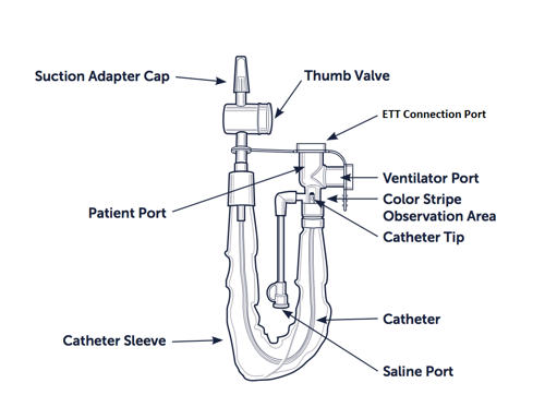
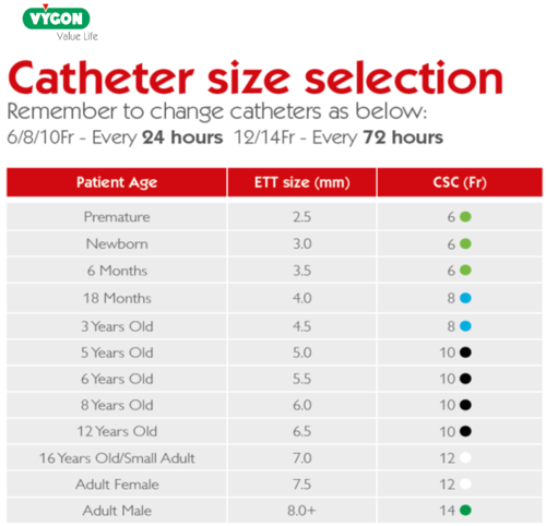
For open suction, suction catheter selection is determined by doubling the internal diameter of the tracheal tube (Morrow and Argent, 2008, Day et al 2002, APCP, 2015), table 1. For close suction circuit, it is dependent on the manufacturer what size is selected for size of Endo-tracheal Tube. In PICU, the current manufacturer of close suction catheter system used is Avanos (See table 2).

Table 1. Open suction catheter size selection

Table image

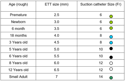
Table 2. Closed Suction catheter (CSC) size selection

Table image

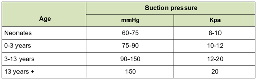
Suction vacuum pressure should be set as low as possible to effectively clear secretions and increasing as required up to a set maximum pressure that is dependent on the age of the child (see table 3).

Table 3. Suction Vacuum pressure

table image

There are no known benefits to performing deep tracheal suction which is related to an increased risk of mucosal trauma and other adverse events (Morrow and Argent 2008, Spence et al 2009, AARC 2010) therefore AARC (2010) advocate shallow suctioning in order to reduce these risks.  Morrow and Argent (2008) recommend that the suction catheter should only be passed to the end of the endo-tracheal tube which can be determined by direct measurement (i.e. closed circuit suctioning). Deep suction should only be performed if the patient is retaining secretions and closed circuit suctioning has not been effective at clearing. Deep suctioning should be kept to a minimum to avoid risk of mucosal trauma. Consider referral to chest physiotherapist if the patient is retaining secretions and requiring deep suction regularly.

It is hypothesized that normal saline instillation may loosen secretions, increase the amount of secretions removed, and aid in the removal of tenacious secretions however there is insufficient evidence to support this hypothesis (AARC 2010).  Caruso et al (2009) believe that normal saline instillation prior to tracheal suction may be associated with decreased incidence of microbiological proven VAP. However, the majority of authors do not consider the instillation of normal saline to be beneficial and may actually be harmful to patients (Edmunds and Scudder 2009, Kuriakose 2008, Rauen et al 2008, Day et al 2002, Morrow and Argent 2008, Halm and Krisko-Hagel 2008, Ridling et al (2003).  Therefore, instillation of normal saline prior to tracheal suction should not be routine practice (AARC 2010, Kuriakose 2008) but may be useful in certain instances or with chest physiotherapy (Paratz and Stockton 2009).  Effective humidification of the ventilator circuit prevents the build-up of thick, tenacious secretions thus reducing the need for instillation of normal saline (Halm and Krisko-Hagel 2008, Ridling et al 2003). 

Procedure – Closed Circuit Endo-Tracheal (ETT) Suction

labelled diagram of the Avanos system

Fig. 1 – Avanos closed suction system

Equipment required for Closed Circuit ETT Suction

  • Stethoscope
  • Gloves
  • Appropriate PPE for procedure
  • Appropriate size of closed circuit catheter for ETT
  • Vacuum suction
  • Suction tubing
  • T-Piece (to be able to hand ventilate patient if required)
  • Leuro Lock syringe
  • 0.9% sodium chloride ampules

Procedure for setting up Closed Circuit ETT Suction

  1. Ensure all equipment required is available.
  2. Select then correct size of catheter required, see table 2. (if the catheter is too large it will occlude the ETT/airway and lead to hypoxia). Ensure the correct size has been chosen.
  3. Wash hands and don appropriate PPE for procedure being carried out.
  4. Attach the closed suction circuit to suction vacuum (Fig 2)
    diagram
  5. Put on Vacuum suction, set at desired pressure, see table 3. Open the thumb valve (Fig 3). Press and hold down the thumb valve to check the check suction is working (Fig 4).
    diagrams 3&4
  6. Attach ventilator port on the closed suction to the patients ETT (Fig 5).
    diagram 5
  7. Lock the thumb valve, to prevent accidental suction being applied when procedure not being carried out (fig 6).  Black catheter tip should be evident in the observation widow when the close suction system is not in
    diagram 6&7
  8. Attach the daily change sticker across the bottom of the thumb control valve. Suction catheters should be changed as per manufacturer’s instructions.

Depth of Catheter Insertion for Closed Circuit ETT Suction

Diagram showing the location of the observation areas

Take note of the last number visible on ETT. When inserting the Closed Suction catheter (CSC) advance until the numbers match. Then advance the CSC another 0.5cm to ensure the catheter has gone just beyond the length of the ETT.

If the numbers on the ETT are not visible for any reason please refer to table 2 which will provide correct depth that the CSC can be advanced to and viewed in the observation area.

Step by Step guide to Closed Circuit ETT suction

Procedure for Close Circuit Endo - Tracheal Suction

Rationale/ Evidence

1) Assess the need for tracheal suction (see indications above):

  • Whenever possible the child should be encouraged to clear their airway by coughing or other clearance techniques.
  • If the child is able to mobilise secretions into their airway independently, closed circuit suction should be performed for secretions within the trachea. Open suction should only be performed when there is evidence of retained secretions which closed circuit suction has failed to clear. Or if indicated during chest physiotherapy.

Suction is an invasive and potentially dangerous procedure that may be traumatic to the child.  It should therefore be used with care and thorough assessment, and when less invasive procedures are ineffective.

The major concerns are the possibility of:

  • Respiratory distress
  • Hypoxia
  • Vomiting and risk of aspiration
  • Tracheobronchial trauma and granulation or ulcer formation
  • Pneumothorax in infants
  • Raised intracranial pressure or arrhythmias if carina touched
  • Intraventricular haemorrhages in premature infants
  • Hypo or hypertension

2) Check all equipment is available and in working order. Visually check for blockages in the internal suction catheter. Ensure the suction tubing is attached to the suction port and suction is switched on.

N.B – CSC should already be in situ when patient is initiated on to mechanical ventilation therapy. If not follow guidance at section 5.1.2 on setting up closed circuit ETT suction.

Suction equipment needs to be prepared and ready to use.

To maintain safety of the person undertaking the procedure.


3) Explain and discuss the procedure with the child and family if possible. Gain consent.


To ensure understanding and consent is given.

Encourage cooperation and reduce anxiety.                                                                      

4) Assessment – Observe the child’s respiratory status and baseline values, e.g. heart rate, blood pressure, work of breathing and colour. 

Note the child’s normal and accepted level of oxygen saturation level pre-suction.

Maintain child’s safety.  To enable assessment of the effectiveness of treatment. 

5) Ensure relevant personnel are present to assist with procedure.

1 qualified person procedure

6) Wash hands and don appropriate PPE for procedure being carried out.

Adhere to infection control policies.

To maintain safety of the persons undertaking the procedure.

7) Unlock system via the thumb valve.          

diagram showing how to use the thumb valve

Vacuum suction should already be on. Test the suction equipment is working by pushing down on the thumb valve.
Using the thumb valve

There is a risk of atelectasis (partial lung collapse) and hypoxia if the suction pressure is too high.

Observe the pressure on the manometer of the suction machine and adjust as required. Values for correct pressure are on Table 2.

N.B - No audible sound of suction will be present due the nature of the closed circuit and this will not cause any determent to the patient as the catheter tip is still within the observation area.

To minimise the risk of atelectasis and hypoxia.      

8) If identified (on an individual clinical basis), pre-oxygenate the child with oxygen suction support on the ventilator prior to suctioning.

To prevent hypoxia.                                    

9) Stabilise the ETT and suction catheter with one hand.
Showing how to stabilise the ETT

With the thumb and forefinger of your dominant hand advance the suction catheter.

N.B – Smooth out the sleeve around the suction catheter when advancing to prevent “bunch up” at the ETT end of the CSC.

To keep the airway secure

10) Advance the suction catheter to the depth required (Table 2), ensuring the correct depth has been achieved by viewing the number on the suction catheter in the observation area.

The catheter tip can cause trauma to the carina if passed recklessly and if suction is applied directly onto the carina, this can also cause trauma.

Patient safety, prevent harm to patient.

11) Once suction is applied hold for two seconds at the desired depth, then continue to apply suction as you pull catheter back from the airway, so that the tip of the catheter is in the observation area. This should be performed slowly to improve yield but no longer than 10s once suction applied.
Using the thumb valve

To maximise yield of suction effort.

To reduce risk of volume loss and hypoxia.

12) Reassess patient – return to section 1 of procedure. If a further suction effort is required, allow time for the patient to recover and provide pre-oxygenation via the ventilator suction support setting if clinically required for the patient.

Allow time to recover to prevent hypoxia and allow recruitment. Change catheter to ensure a clean technique and reduce infection risk.

13) Prepare an ampule of 0.9% sodium chloride or leur slip syringe. Open saline port and attach ampule.  Ensure the catheter tip is visible within the observation area.
How to attach ampule to saline port

Use 0.9% Sodium chloride to flush and clear the system before use on the patient.

To enable early recognition of any potential complications pre suction.

To maintain the child’s safety.

Pushing through saline

Push in 0.9% sodium chloride whilst pressing on the thumb valve to clear the catheter before use. The black catheter tip should remain in the observation widow at this time.

To ensure patency of the suction catheter before use.

15) Ensure the catheter tip is in the observation window.
Checking observation window

Lock the thumb valve.
Locking the thumb valve

To maintain the child’s safety.

16) If secretions are thick, sticky or non-yielding consider referral to chest physiotherapists or medical staff.

Try to avoid disconnecting the CSC system for open suction unless the patient’s condition deteriorates.

Maintain patient safety.

Chest physiotherapist provide treatment techniques that aid with the removal of secretions and improve airway clearance.

Saline nebulisers can aid in loosening thick, sticky secretions for airway clearance.

Adapted from ACPC, 2015

Procedure – Open Endo Tracheal (ET) Suction

Equipment required for Open ET suction

  • Stethoscope
  • Gloves ( box of)
  • Measuring tape
  • Appropriate PPE for procedure
  • Appropriate size of suction catheter
  • Vacuum suction
  • Suction tubing
  • T-Piece (hand bag ventilation)
  • Oxygen point
  • Orange disposable bag or orange bin close by

Step by Step Guide to Open ET suction

Procedure

Rationale/ Evidence

1) Assess the need for tracheal suction (see indications above):

  • Whenever possible the child should be encouraged to clear their airway by coughing or other clearance techniques.
  • If the child is able to mobilise secretions into their airway independently, closed circuit suction should be performed for secretions within the trachea.

Open suction should only be performed when there is evidence of retained secretions which closed circuit suction has failed to clear. Or if indicated during chest physiotherapy.

Suction is an invasive and potentially dangerous procedure that may be traumatic to the child.  It should therefore be used with care and thorough assessment, and when less invasive procedures are ineffective.

The major concerns are the possibility of:

  • Respiratory distress
  • Hypoxia
  • Vomiting and risk of aspiration
  • Tracheobronchial trauma and granulation or ulcer formation
  • Pneumothorax in infants
  • Raised intracranial pressure
  • arrhythmias if carina touched
  • Intraventricular haemorrhages in premature infants
  • Hypo or hypertension

2) Check all equipment is available and in working order for open ET suction, as per section above.

Suction equipment needs to be prepared and ready to use.

To maintain safety of the person undertaking the procedure.                                                   

3) Ensure the correct size of suction catheters are available and depth of suction has been ascertained for the correct size of ETT (Table 1 and appendix 1).

N.B – suction catheter length should be measure with a tape measure before suction carried out.

Also have some smaller sizes of catheter in case issues advancing appropriate size for ETT.

Suction can cause mucosal trauma and arrhythmias (abnormal heart rhythm).

Using a larger than required catheter can cause mucosal or airway trauma and not necessarily improve yield.

The catheter tip can cause trauma to the carina if passed recklessly and if suction is applied directly onto the carina, this can also cause trauma.

Maintain patient safety and prevent harm.

4) Explain and discuss the procedure with the child and family if possible.  Gain consent.

To ensure understanding and consent is given.

Encourage cooperation and reduce anxiety.                                          

 5) Assess and observe the child’s respiratory status and baseline values, e.g. heart rate, blood pressure, work of breathing and colour.  Note the child’s normal and accepted level of oxygen saturation level pre-suction.

Maintain child’s safety. 

To enable assessment of the effectiveness of treatment. 


6) Ensure relevant personnel are present to assist with procedure.

2 qualified person procedure.

7) Wash hands and don appropriate PPE for the procedure which is aerosol generating.

N.B - Safety goggles are advised if there is a risk of contamination to the person who is undertaking the procedure.

Adhere to infection control policies.

To maintain safety of the person undertaking the procedure.

8) Open and attach the suction catheter to the suction tubing:

  • peel back the catheter to expose the hard plastic connector
  • leave the rest of the catheter in the protective cover

 To ensure sterility of the suction catheter.

9) Switch on the suction machine.  With a gloved hand, test the suction equipment by placing the thumb over the end of the suction tubing for 5-10 seconds.

Observe the pressure on the manometer of the suction machine and adjust as required (Table 3).

There is a risk of atelectasis (partial lung collapse) and hypoxia if the suction pressure is too high.                                      

To minimise the risk of atelectasis and hypoxia.

10) If required pre oxygenate the patient with the suction support setting on the ventilator.

To prevent hypoxia.

11) Remove gloves and replace with a new clean set and remove the suction catheter from the package. Hold the end of the suction catheter with dominate and the plastic connector with the other.

Do not touch anything EXCEPT the sterile part of the catheter. 

 To maintain an aseptic non-touch technique.

12) Check the child’s observations and breathing pattern immediately prior to inserting the suction catheter

To enable early recognition of any potential complications. 

To maintain the child’s safety.

13) 2nd personnel should disconnect the ETT from the ventilator and stabilize the ETT. 1st personnel will pass suction catheter into artificial airway to the desired depth.

To maintain an aseptic non-touch technique of the person performing the suction procedure.

14) Depth of suction – the suction catheter should be passed slowly until desired depth has been achieved. (this should have been measured prior to starting procedure with a tape measure).

The catheter tip can cause trauma to the carina if passed recklessly or blindly without a measured indicator of depth. If suction is applied directly onto the carina, this can also cause trauma, swelling and bleeding.

15) Do not rotate the catheter. Once suction is applied hold and apply for around 2 seconds and continue to apply suction as you pull catheter back from the airway. This should be performed slowly to improve yield but no longer than 10s once suction applied.

To maximise yield of suction effort.

To reduce risk of volume loss and hypoxia.

16) 2nd personnel should either reconnect the patient to the ventilator and provide oxygenation via the suction support setting on the ventilator if required. Or if significant desaturation of the patient during the suction procedure attach the patient to the T-Piece and deliver hand ventilation until the patient recovers.

Prevent hypoxia.

17) Reassess patient – return to section 1 of procedure, if a further suction is required. Allow the patient to recover.

Change the suction catheter for each suction attempt and renew dominant hand glove.

If no further suction is required reconnect the patient back to mechanical ventilation and observe the patient post suction.

Allow time to recover to prevent hypoxia and allow recruitment.

Change catheter to ensure a clean technique and reduce infection risk.

16) If secretions thick, sticky or non yielding consider referral to chest physiotherapists or medical staff.

Try to avoid disconnecting the CSC system for open suction unless the patients condition deteriorates.

Maintain patient safety.

Chest physiotherapist provide treatment techniques that aid with the removal of secretions and improve airway clearance.

Saline nebulisers can aid in loosening thick, sticky secretions for airway clearance.

Adapted from ACPC, 2015.

Evidence

This SOP has been constructed following review of sourced textbooks and a literature search of Medline, CINAHL and Cochrane databases.  No national guidelines are available on this topic so international guidelines were utilized and other trust’s guidelines were accessed.  Internal and external expert review and opinion were also taken into consideration.  Physiotherapists, nurse educators, senior nursing staff and Intensivists were all consulted, therefore, the best available evidence was used to construct this SOP and the recommendations it makes. 

Review

There will be a review of this SOP every three years from the date of approval.

Contact/support information

For further assistance contact Senior nursing staff on duty: phone 84713

Appendix 1: ETT Closed Suction Catheter size selection chart

Image of Catheter Size Selection Chart

Editorial Information

Last reviewed: 10/06/2024

Next review date: 31/01/2027

Author(s): M Chalmers, L Montgomery, R Kennedy. Adapted by L Moore (PICU Clinical Nurse Educator), R. Marscheider (PICU Physiotherapist).

Version: 2.1.2

Author email(s): ross.marscheider@ggc.scot.nhs.uk, lorraine.moore2@ggc.scot.nhs.uk.

Approved By: PICU Consultant Group

References
  1. AARC (2010) “Endotracheal suction of mechanically ventilated patients with artificial airways”. Respiratory Care. Vol. 55, No.6, pp.758-764

  2. Caruso, P., Denari, S., Soraia, A.L., Ruiz, A.L., Demarzo, S.E., Deheinzelin, D. (2009) “Saline instillation before tracheal suctioning decreases the incidence of ventilator associated pneumonia”.  Crit Care Med. Vol 37, No 1, pp.32-38

  3. Day, T., Farnell, S., Wilson-Barnett, J. (2002) “Suctioning: A review of current research recommendations”. Intensive And Critical Care Nursing.  Vol. 18, pp. 79-89

  4. Edmunds, M.W. And Scudder, M.S., (2009) “Bringing evidence to the process of endotracheal suctioning”. Intensive Crit Care Nurse, Vol.25, pp. 21-30

  5. Halm, M.A., Krisko-Hagel, K. (2008) “Instilling normal saline with suctioning: beneficial technique or potentially harmful sacred cow?”  American Journal Of Critical Care.  Vol 17, No. 5, pp. 469-472

  6. Kuriakose, A. (2008) “Using the Synergy Model as best practice in endotracheal tube suctioning of critically ill patients”.  Dimensions Of Critical Care Nursing.  Vol. 27, No. 1, pp. 10-15

  7. Morrow, B., Futter, M., Argent, A. (2006) “Effect of endotracheal suction on lung dynamics in mechanically-ventilated paediatric patients.”. Australian Journal Of Physiotherapy.  Vol 52, pp. 121-126

  8. Morrow, B.M., Argent, A.C. (2008) “A comprehensive review of pediatric endotracheal suctioning: Effects, indications, and clinical practice.” Paediatric Crit Care Med. Vol. 9, No. 5, pp.465-477

  9. Paratz, J.D, Stockton, K.A. (2009) “Efficacy and safety of normal saline instillation: a systematic review."   Vol. 95, pp. 241-250

  10. Pritchard, M.A., Flenady, V., Woodgate, P.G. (2010) “Preoxygenation for tracheal suctioning in intubated, ventilated newborn infants.” Cochrane Database of Systematic Reviews. 

  11. Rauen, C.A., Chulay, M., Bridges, E., Vollman, K.M., Arbour, R. (2008) “Seven evidence-based practice habits: putting some sacred cows out to pasture.” Critical Care Nurse.  Vol. 28, No. 2, pp. 98-124

  12. Reiger, H., Kuhle, S., Ipsiroglu, O.S., Heinzl, H., Popow, C.N. (2005) “Effects of open vs. closed system endotracheal suctioning on cerebral blood flow velocities in mechanically ventilated extremely low birth weight infants.” J Perinat Med. 33(5):435-41.

  13. Ridling, D.A., Martin, L.D., Bratton, S.L. (2003) “Endotracheal suctioning with or without instillation of isotonic sodium chloride solution in critically ill children." American Journal Of Critical Care. 12, No.3, pp. 212-219

  14. Rychik, J., Bush, D.M, Spray, T.L., Gaynor, W., Wernovsky, G. (2000) “Assessment of pulmonary/systemic blood flow ratio after first-stage palliation for hypoplastic left heart syndrome: development of a new index with the use of doppler echocardiography.” The Journal Of Thoracic And Cardiovascular Surgery.  Vol. 120, pp. 81-87

  15. Spence, K., Gillies, D., Waterworth, L. (2009) “Deep versus shallow suction of endotracheal tubes in ventilated neonates and young infants”. Cochrane Database of Systematic Reviews.

  16. Stokowski, L.A. (2009) “Preventing ventilator associated pneumonia in infants and children” Medscape.

  17. Trevisanuto, D., Doglioni, N., Zanardo, V. (2009) “The management of endotracheal tubes and nasal cannulae: the role of nurses.”  Early Human Development.  Vol. 85, pp. 85-87

  18. Association For Paediatric Chartered Physiotherapists (2020) Guidelines For Nasopharangeal Suction Of A Child Or Young Adult.

  19. Maggiore, S. M., Iacobone, E., Zito, G., Conti, M., Antonelli, M., Proietti, R. (2002). Closed Versus Open Suctioning Techniques. Minerva Anestesiol Vol 68:360-4.