exp date isn't null, but text field is
This guideline has been written to standardise the measurement of intra-abdominal pressure and it describes which high risk patients this should be measured in.
This guideline is intended to be used by healthcare professionals involved in the care of children at risk of raised intra-abdominal pressure, especially in the paediatric critical care uint. It describes how intra-abdominal pressure can be measured using a urinary catheter and a pressure transducer.
Nursing & medical staff especially within PICU
The abdomen can be defined as a closed space or compartment enclosed by the spine, pelvis, diaphragm and abdominal wall. The elasticity of the walls and character of the contents of the abdomen, determine the pressure within it at a given time. The Intra-abdominal pressure is defined as the steady state pressure concealed within the abdominal cavity [1].
Abdominal pressure therefore varies depending on the physiological status of the patient. Abdominal pressure increases with inspiration, use of abdominal muscles, and increasing volume of fluid (eg. ascites, blood) in the abdominal space. It also increases with visceral expansion or collection within the viscera whether that is air, fluid or faeces. Intra-abdominal pressure is also affected by conditions which limit abdominal expansion such as 3rd space oedema or burn eschars.
Normal intra abdominal pressure may range from subatmospheric to 0mmHg. In the critically ill, IAP is frequently elevated above normal. In critically ill adults normal IAP is defined as 5-7mmHg [1].
In one group of children where IAP was measured directly via peritoneal dialysis catheter post cardiac surgery, median IAP was found to be 4mmHg with a range of 1-8mmHg [3].
Similar to cerebral perfusion pressure, abdominal perfusion pressure is defined as the mean arterial pressure minus the intra abdominal pressure [1]. It has been accepted in adults as a more accurate predictor of visceral perfusion than MAP or IAP alone. Again, in adults a target APP of >60mmHg has been shown to correlate with better outcomes, though this has not been studied in children.
Definition: The World Society of the Abdominal Compartment Syndrome defines ACS as sustained intra-abdominal pressure of ≥20mmHg with new organ dysfunction or failure [1].
Abdominal compartment syndrome represents the pathophysiological consequences of a raised IAP, including effects on lung compliance and ventilation, a critical reduction in the perfusion of the intra-abdominal organs leading to oliguria, renal impairment and splanchnic ischaemia. Increased intracranial pressure has also been reported.
ACS may be classified as primary or secondary. Primary ACS is characterised by acute ACS occurring as a result of an intra-abdominal cause such as abdominal trauma, peritonitis or retroperitoneal haemorrhage.
Secondary ACS is characterised by the presence of subacute or chronic IAH that develops as a result of an extra-abdominal cause such as sepsis, capillary leak, major burns or other conditions requiring massive fluid resuscitation.
In one of the few paediatric studies of outcomes of children with ACS [4] in a mechanically ventilated subset of patients admitted to PICU, 4.7% (14/294) developed ACS. Of those, there was a 50% mortality among those with ACS vs 8.4% of those who did not develop ACS.
Intra-abdominal pressure |
|
Normal |
5-7 mmHg |
Grade I |
12-15 mmHg |
Grade II |
16-20 mmHg |
Grade III |
20-25 mmHg |
Grade IV |
>25 mmHg |
Abdominal examination and clinical judgement are inaccurate methods of measuring abdominal pressure. A number of methods have been used in order to quantify abdominal pressure in the past including intra-gastric, rectal, intra-uterine and direct abdominal pressure monitoring [3].
The ‘gold standard’ measurement of abdominal pressure monitoring is now widely accepted to be the intravesical method [2,3,5,6]. It has been found to be the method which most closely correlates to direct intra-abdominal pressure when measured by intra–peritoneal catheter in children when instilling a volume of 1 ml/kg of fluid into the bladder [3].
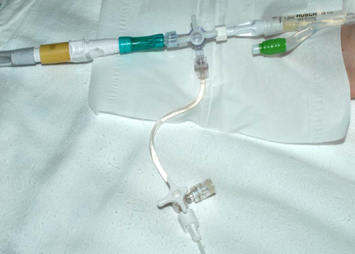
Fully set up it should look like this:
Positioning – The WSACS has recommended that the measurement of abdominal pressure should be measured in the following position and time:
Procedure
The world congress on ACS has proposed the following risk factors for IAH/ACS [2].
Decreased abdominal wall compliance
Increased intra-abdominal contents
Capillary Leak /Fluid Resuscitation
The WSACS suggests we should assess patients with potential IAH according to the following algorithm:
Current evidence surrounding management of IAH and ACS on which to base best practice guidelines in adults are mainly retrospective [2] and include small studies based on specific groups of patients – ie. post-operative abdominal surgical or burns patients [2].
The evidence base in children is even more scant and appears to be currently limited to case reports. For this reason, management of any child with raised abdominal pressure should be discussed with the consultant on call.
The WSACS has published an algorithm for management of ACS based on best available current evidence in adults, which is expected to change as we gain more knowledge on the subject. For interest only it can be found online by following this link:
The medical management algorithm has been recently updated (1/2/09):
IAH ACS medical management 2014
Medical treatment options for use in the first instance are:
Reduction in abdominal wall compliance
Evacuate Intraluminal Abdominal Contents
Evacuate Extraluminal Abdominal Collections
Correct Positive fluid balance
Surgical treatment options
The WSACS recommends consideration of early abdominal decompression in primary ACS. In secondary ACS, when all medical treatment options have failed to relieve IAH, abdominal decompression with temporary abdominal closure is recommended [2].
Last reviewed: 01 November 2012
Next review: 01 November 2014
Author(s): A McGettrick