Eye Care in PICU
Search RHCG Website
Select your language

Eye Care in PICU

exp date isn't null, but text field is

Objectives

  • To protect the eye in unconscious and ventilated patients within PICU
  • Identify ocular disease and those requiring referral
  • Management of minor eye disease

Scope

This guideline is intended for healthcare professionals caring for patients within the Paediatric Intensive Care Unit at the Royal Hospital for Children, Glasgow.

Whilst ocular conditions may alter between different ages, this basic ocular management guidance applies for all children from neonates to teenagers. There are some important age related considerations at the end of the guideline

Audience

Medical, nursing and allied professionals caring for patients in PICU.

Ophthalmology contact details

Most inpatient referrals can be sent via trakcare:

  • Select new request, other,
  • under item select “Ophthalmology Inpatient referral – Paediatric” and fill in the required details.

For more urgent referrals, or for advice, please contact 85808 Monday to Friday 9am – 5pm, except on Wednesday afternoons. For Wednesday after 1pm or out of hours, please contact ophthalmology on-call via switchboard.

Introduction

Patients in intensive care are at risk of developing corneal defects as normal ocular defence mechanisms are challenged [1-7]. Sedatives and paralysis interfere with eyelid closure [8]. Passive closure may occur, but is often incomplete [1]. Lack of blinking fails to renew the tear film leading to the potential retention of micro-organisms. Mechanical ventilation encourages venous stasis and fluid retention, leading to conjunctival chemosis [2]. Capillary leak and fluid shifts associated with sepsis similarly cause conjunctival chemosis [9]. The exposed cornea becomes dry, and small defects may develop [3]. Keratitis occurs if micro-organisms adhere to the damaged corneal surface [10,11]. Corneal defects are painful and a source of agitation, as well as being potentially blinding.

The proposed algorithm uses eyelid position and whether the patient is lying prone or not to guide treatment and been made considering guidance from the Royal College of Ophthalmologists [12]. A randomized controlled trial comparing ocular lubricant to passive eye closure, found lubricant to be favourable [1]. When lubricant was compared to eye taping or covering there was no significant difference between the treatments [13].

We have selected lubricants with/without taping as treatments in the first instance, as these are easy to use, effective and cost-efficient. Complications of this form of management are expected to be minimal. Temporary blurred vision may occur with the use of ointments. As well as protection of the cornea in at risk patients, these guidelines also discuss microbial conjunctivitis and keratitis, chemosis and the role of ophthalmology review in patients with positive fungal cultures.

Protective measures in unconscious or ventilated patients (algorithm)

Eye care algorithm for children who are intubated and ventilated

Protective measures in unconscious or ventilated patients (procedures)
Application of eye ointment

Pull down the lower eyelid and apply a small amount of eye ointment to the inside of the eyelid and manually close in poor lid closure to ensure ointment is spread over the eye. The ointment should not be applied over closed eyelids.

Ensure a different tube is used for each eye. Lubricants used should always be ointments as these last longer than drops. Hylonight should be used in the first instance, however if this is not available then carbomer gel or simple eye ointment can be used.  

If drops are also required, use these prior to ointment. Wait 5 minutes between drops if multiple drops are required.

If chloramphenicol antibiotic ointment is being used, this works well as a lubricant so often the lubricant can be omitted at times where this is also being used.


Eyelid bathing

Use 0.9% saline and ensure the lids are closed. Use a new piece of gauze and ensure this edge is not above the lid margin (lower lid) or below it (upper lid). Ensure the gauze swab goes from the inner to the outer canthus and dry it in the same manner.


Closing the eyelids

This may be done via the following methods:

Manual closure:

Eyelid taping
This may cause eyelid injury or irritation and can be distressing to relatives so should only be undertaken where necessary. After applying lubricant, close the lid and stick the tape (such as Micropore) onto the lid and lashes horizontally to ensure closure.

Cling film
Apply a 10cm x 10cm square over each eye and change it every shift. It does not cause damage if in contact with the eyeball.

Hydrogel or silicone dressings/pads
Use with care to avoid damage to the eye, eg Kerrapro, Gelliperm may be used instead of taping if oedema prevents manual lid closure. They should be changed at least once per shift or sooner if drying out (curling edges).


Proned patients

These patients are more at risk due to compression and conjunctival swelling (chemosis). Lubricants and taping should be used (see algorithm) and a 3 pin head holder can also be used to avoid compression, as in spinal surgery.

The above eye care regime may be discontinued once the patient is awake, eyelid movement is re-established and extubation is performed.

Ocular pathology within PICU
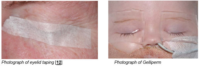
Exposure keratopathy

This is a corneal epithelial defect which occurs in those with incomplete lid closure. It is best seen with fluorescein eye drops and a blue light preferably, but may also be visible with a white light.

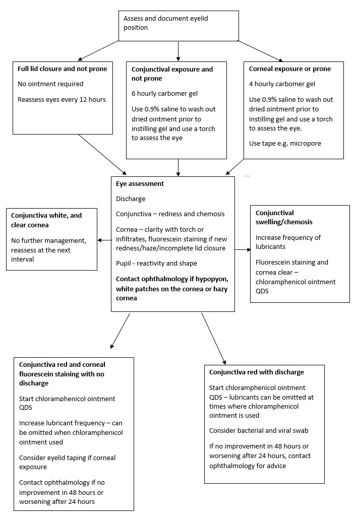
In the first instance, use chloramphenicol ointment 4 times a day, increase the frequency of lubricants and consider lid taping.

Contact ophthalmology if:

  • The cornea is not clear with a bright light
  • White patches are present on the cornea
  • The epithelial defect covers more than one third of the cornea
  • There is no improvement within 48 hours after increasing management

If there is a corneal abrasion with full lid closure, chloramphenicol 4 times a day for 5-7 days can be used.
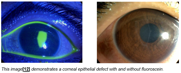
 

Chemosis

Conjunctival chemosis, causing swelling and bulging of the conjunctiva, is common in patients who are ventilated, particularly with positive pressure ventilation, patients with generalised oedema or those that are prone.

Chemosis can lead to poor lid closure leading to exposure of the cornea and infection. These patients can often be managed with increased lubrication and taping however refer to ophthalmology if:

  • The cornea is not clear with a bright light
  • White patches are present on the cornea
  • The epithelial defect covers more than one third of the cornea
  • There is no improvement within 48 hours after increasing management


Microbial Infections

Respiratory secretions may be a common source of bacteria in those in PICU, so care should be given when suctioning is being performed. This should be done from the side rather than at the head, and the eyes should be covered.

Conjunctivitis

This is an infection of the conjunctiva.The eye is usually sticky with debris on the lashes and a red eye. If the eye is red without stickiness other causes should be considered. Discuss with ophthalmology if the cornea is not clear.

Conjunctivitis may be bacterial or viral. Use chloramphenicol ointment 4 times a day for 7 days and send a viral and bacterial swab. If the swab shows sensitivities to other antibiotics but there is a clinical improvement, continue chloramphenicol.

Microbial keratitis

This is an infection of the cornea. Patients with exposure keratopathy are more vulnerable to this due to loss of the protective epithelial barrier. A white patch may be visible on the cornea which usually stains with fluorescein, the cornea may be hazy with a bright torch and the eye will turn red. These patients should be referred to ophthalmology.

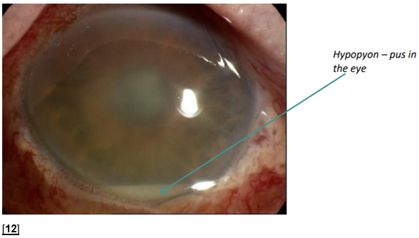
Endogenous endophthalmitis

This is a serious and rare complication of a systemic infection.The eye may be red with poor visibility of the pupil and iris with a bright torch. It should be suspected in those with a white line of pus called a hypopyon andrequires an urgent ophthalmology review. It indicates active sepsis and requires systemic treatment.


Chemical eye injury
  • 1 litre of saline irrigation
  • pH strip – test conjunctiva to ensure pH is 7-8, if not continue irrigation until pH normalises
  • Fluoroscein – if there is fluoroscein staining use chloramphenicol ointment 4 tmes a day for 5 days
  • Contact ophthalmology for advice if the substance is significantly acidic or alkali
Ocular examination in systemic fungal infection

Endogenous ocular involvement from candidaemia is rare but can be sight threatening.

Recent published literature suggests a decreasing prevalence of ocular involvement in recent years, possibly due to earlier initiation of systemic therapy, and greater intraocular penetrance of newer generation antifungals.A recent local study in the adult sector of NHS GG&C suggests a prevalence of 1.3%. [14]. This study found that no change in treatment was required as a result of positive ocular findings.

There is no published literature demonstrating ocular candidiasis in the absence of positive blood cultures.

Guidance

The Royal College of Ophthalmologists guidelines [12] from 2020 suggests that routine screening of fungal culture positive patients is not indicated and should only be performed as an exception on a case by case basis, taking these principles into account:

No examination required: awake and asymptomatic

May need examination, refer to ophthalmology for advice: awake and symptomatic, unable to report symptoms

Must be examined: Very abnormal eye appearance eg hypopyon, cloudy pupil, possible ocular perforation etc.

Considerations to allow whether examination is required, and timing of the examination should be performed based on:

  • Risk, prognosis, microbiology results, ability of current/planned treatment to penetrate the eye, patient position, ability to examine.

There is no indication for ophthalmology assessment of patients with no positive blood culture evidence of candida or other fungi, in the absence of other ocular symptoms or signs. Patients with positive fungal line tip cultures do not require ophthalmology review if they are asymptomatic – if there is concern blood cultures should be carried out.

Age related considerations

Chloramphenicol 0.5% drops are contraindicated in patients under 2 (theoretical fertility issues related to boric acid) [15]. Chloramphenicol 1% ointment does not contain boric acid so is safe to use in this age group.

Under the age of 7, sensory deprivation can irreparably damage vision causing amblyopia. In PICU, maintaining the integrity of the corneal surface to prevent corneal scarring in this age group is a priority. Sensory deprivation may be reversible.

References
  1. Lenart SB, Garrity JA. Eye care for patients receiving neuromuscular blocking agents or propofol during mechanical ventilation. Am J Crit Care 2000; 9 (3): 188-91.
  2. Dua HS. Bacterial keratitis in the critically ill and comatose patient [letter]. Lancet 1998; 351: 387.
  3. So HM, Lee CCH, Leung AKH, Lim JMJAL, Chan CSC, et al. Comparing the effectiveness of polyethylene covers (GladwrapTM) with lanolin (DuratearsR) eye ointment to prevent corneal abrasions in critically ill patients: A randomised controlled study. Int J Nurs Stud 2008; 45: 1565-71.
  4. Germano EM, Mello MJG, Sena DF, Correia JB, Amorim MMR. Incidence and risk factors of corneal epithelial defects in mechanically ventilated children. Crit Care Med 2009; 37 (3): 1097-1100.
  5. Imanaka H, Taenaka N, Nakamura J, Aoyama K, Hosotani H. Ocular surface disorders in the critically ill. Anesth Analg 1997; 85: 343-6.
  6. Hernandez EV, Mannis MJ. Superficial keratopathy in intensive care patients. Am J Ophthalmol 1997; 124 (2): 212-6.
  7. Mercieca F, Suresh P, Morton A, Tollo A. Ocular surface disease in intensive care patients. Eye 1999; 13: 231-6.
  8. Sorce LR, Hamilton SM, Gauvreau K, Mets MB, Hunter DG, et al. Preventing corneal abrasions in critically ill children receiving neuromuscular blockade: A randomised controlled trial. Paediatr Crit Care Med 2009; 10 (2): 171-5.
  9. Ezra DG, Chan MPY, Solebo L, Malik AP, Crane E, et al. Randomised trial comparing ocular lubricants and polyacrylamide hydrogel dressings in the prevention of exposure keratopathy in the critically ill. Intensive Care Med 2009; 35: 455-61.
  10. McClellan KA. Mucosal defense of the outer eye. Surv Ophthalmol 1997; 42: 233-46.
  11. Parkin B, Turner A, Moore E, Cook S. Bacterial keratitis in the critically ill. Br J Ophthalmol 1997; 81: 1060-3.
  12. Royal College of Ophthalmologists (RCOphth). Eye Care in the Intensive Care Unit (ICU). 2020. 
  13. Bates J, Dwyer L, O’Toole L, Kevin L, O’Hegarty N, et al. Corneal protection in critically ill patients: a randomised controlled trial of three methods. Clin Intensive Care 2004; 15 (1): 23-6.
  14. El-Abiary, Jones B, Williiams G, Lockington D. Fundoscopy screening for intraocular candida in patients with positive blood cultures—is it justified? Eye 2018 32:1697-1702.
  15. Royal College of Ophthalmologists (RCOphth). Safety Alert: Boron additives in Chloramphenicol drops; should ophthalmologists be concerned? 2021. 
Editorial Information

Last reviewed: 01 March 2022

Next review: 31 March 2025

Author(s): Chloe Shipton, Eoghan Millar, Sonul Gajree, Manaim Shah, Graham Bell

Author Email(s): eoghan.millar2@ggc.scot.nhs.uk

Co-Author(s): Based on the previous version from 2011 by Karen McCall, Jeanette Grady, Mangosha Hussin, Julie Richardson, Gordon Dutton