In addition to routine Cranial USS imaging, as described above, all premature infants should have measurements of their OFC performed weekly to identify cases of Post-haemorrhagic Ventricular Dilatation (PVHD). All babies with PHVD need regular scans to monitor the progression of the ventricular dilatation and to assess the possible requirement for ventricular drainage. A number of measures have been described to monitor ventricular dilatation the most widely used being the ventriculo-cranial ratio (VCR) and the ventricular index (VI). There is no clear advantage to either measure but it is important to use the same measurement in an individual patient to monitor change over time.
Ventriculo-cranial ratio (VCR)
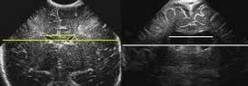
Measurement of the ventricular system needs to be performed on a symmetrical, easily reproducible view. The ventriculo-cranial ratio (VCR) is the ratio of distance between the lateral sides of the ventricles and the biparietal diameter. This is usually expressed as a percentage with a normal value of around 33-36% in a preterm infant. This value is of most use in monitoring the degree of change between successive measurements. An increasing VCR should trigger frequent reassessments with measurements of the cerebral resistive index (see below).
Ventricular Index (VI)
The Ventricular index, as described by Levine, is the absolute distance between the falx and the lateral wall of the anterior horn in the coronal plane at the level of the third ventricle. Values more than 4mm above the 97th centile for gestational age are indicative of significant ventricular dilatation (see chart). A study by Brouwer et al compared early CSF drainage, at a ventricular index of 4 mm above the 97th centile, to drainage beyond this threshold, and showed that early treatment was associated with better development quotient at 2 years and a decreased likelihood of shunt dependence; there was, however, no reduction in the rate of cerebral palsy with early drainage.
Resistive Index (RI)
In 1976, Pourcelot introduced the concept of RI, which is calculated by the following formula: RI = (S-D)/S where S and D stand for systolic and diastolic velocities measured in the cerebral arteries. The infant’s blood pressure and carbon dioxide tension need to be taken into account when measuring the RI as these can affect cerebral blood flow. Variations in the RI demonstrate that cerebral blood flow is not well regulated and are often associated with adverse outcomes. It is important to interpret the RI with caution as cerebral blood flow is always changing due to haemodynamic alterations and there is also considerable inter and intra observer variation.

Interpretation
High RI
A high RI (>0.85) corresponds to low blood flow velocity where vascular resistance is high (eg hydrocephalus). Infants with values higher than this may require ventricular drainage to reduce intracranial pressure.
NB: A high RI must be interpreted with caution in an infant with a PDA as these infants may have a low diastolic velocity due to ductal steal. This will give a high RI value even in infants with normal intracranial pressure.
Low RI
A low Resistive Index corresponds to high blood flow velocity where vascular resistance is low (eg HIE). In normothermic infants, an RI of <0.55 has a positive predictive value for poor neurological outcome of 84% (95% CI 73%, 91%). However in cooled infants the positive predictive value is lower - 60% (95% CI 45%, 74%). (Elstad M et. al,)

 GGC Paediatric Guidelines