Search RHCG Website
Select your language

Patent ductus arteriosus (PDA) : surgical ligation

exp date isn't null, but text field is

Objectives

Paediatric cardiothoracic surgical services for Scotland are centralised at the Royal Hospital for Children (RHC), Glasgow. Therefore all patent ductus arteriosus (PDA) ligations in premature infants in Scotland are undertaken at RHC Glasgow, usually in the Neonatal Intensive Care Unit (NICU). 

This guideline outlines the potential indications, referral pathway, assessment processes and perioperative protocol for PDA ligation of the premature infant in NICU, RHC. The guideline is intended for the use of referring neonatologists, nursing and medical staff in NICU, paediatric cardiologists and members of the theatre, anaesthetic and cardiothoracic team at RHC, Glasgow.

Scope

This guidance is principally designed for premature infants, typically born at <28 weeks gestation, who have a high risk associated morbidity with persistent PDA and whose surgery, if ligation is required, should be undertaken in NICU. However more mature infants can develop heart failure associated with persistent PDA and the same principals or referral and assessment apply.

This protocol does not apply to infants referred to cardiology for PDA closure by cardiac catheterisation. Currently, infants being considered for trans-catheter PDA closure should weigh 4 kg although with the use of venous occlusion devices this may fall in the future.

Background

PDA management in preterm infants: general                                                            

An unrestrictive PDA in a premature infants can cause: progressive cardiac failure, prolonged ventilator dependence and can contribute to chronic lung disease (1,2). Additionally, associations between feed intolerance, necrotising enterocolitis, hypotension (3) and cerebral perfusion (4) are established. There is considerable animal evidence that PDA adversely affects lung compliance (5,6).The earliest (1970s) clinical trials demonstrated a reduction in ventilator support and hospital stays following surgical ligation(7). With improvements in perinatal care the optimal management of the PDA has been more controversial(8) but whilst there is no place for universal prophylactic PDA closure, medically or surgically, PDA ligation still has a role for preterm infants who (with) have persistent moderate/large PDA, who develop cardiac failure and who are not responsive to medical therapy.

Despite 50 years of clinical trials there is no clear picture or national/ international guidance on the management of PDA. After the earliest clinical trials, most randomised controlled trials (RCTs) either focussed on refinement of treatment or prophylactic treatment to reduce periventricular haemorrhage and improve developmental outcomes. Recent Cochrane reviews (9,10) of the more recent “treatment versus no treatment “demonstrate reductions in hospital stays but no other differences in outcome mortality of short term outcomes. Apparent negative results of trials have resulted in a therapeutic nihilism and a drift away from treatment. However the Cochrane authors acknowledge the heterogeneity of gestational age (GA) of infants included, potential selection bias and the need for appropriately targeted and powered trials. Despite this, PDA management (primarily medical but also surgical in selected populations) remains a standard in many institutions with the best neonatal outcomes. Evidence to support this approach comes from quality improvement studies strongly suggesting that moving away from treating PDA results in increasing rates of BPD and more vasopressor treatment of hypotension in the first 2 weeks of life(11,12). Sub analysis of the prophylaxis studies report reduced rates of PVL and better later developmental outcomes with prophylactic indomethacin (13) and MRI brain studies demonstrate lower brain volumes in infants with prolonged PDA exposure (14). Most recently critical appraisal of recent RCTs highlights some of the challenges with subjecting PDA management to RCTs. Problems include wide variation in the selected study population, marked variation in shunt size, shunt exposure, problems with patient selection bias, treatment contamination of the control arms and relatively poor efficacy of medical interventions. Analysis of the most recent RCTs demonstrated only a 20% difference in PDA exposure between the treatment arms in some RCTs (15). In these circumstances analysis of the most recent medical RCTs (with negative findings) demonstrate a strong relationship between the duration of PDA exposure, ventilation and the development of severe grades of BPD (16,17,18,19)

PDA Ligation in preterm infants:

Evidence and outcomes

The situation for surgical ligation of the PDA is more complex. Since the USA National collaborative trial there have been no randomized clinical trials comparing surgical ligation to medical or no PDA treatment (20).

In addition to potential  direct surgical complications (pneumothorax, Post Ligation Impaired LV contractility (PLIC), vocal cord (nerve) palsy) there are concerns that very early handling the preterm lung may also have unexpected adverse effects and increase the risk of BPD (21) so that  that whilst prophylactic PDA ligation decreases NEC rates (22) it may increase BPD(23). However whilst follow up studies have demonstrated higher incidences of BPD and neurosensory impairment (24) a more recent report shows where appropriate risk adjustment is undertaken for case mix babies requiring PDA ligation the increased rates of adverse outcomes do not persist (25).

What is known is that surgical ligation, in contrast to medical therapy, definitively closes the PDA. It has been demonstrated to improve lung compliance (5), may reduce the incidence of NEC (22), is associated in the past and present era with success in getting patients off ventilators (26,27) and, after potential transient decline, improves cerebral perfusion (28).

The approach around the world varies widely. Brooks et al, in 2005, reported an increase in mortality in infants < 28 weeks with PDA not responsive to medical management after losing a PDA ligation service (29). A very recent series from South Korea (median GA 26 weeks) reported that, in infants not responsive to indomethacin, if ligation was deferred from the first week of life until after 10 days of life outcomes were poorer.  Earlier ligation was associated with reduced ventilator requirement and NEC (30). The overall mortality rate for this population was impressively low at 1-2 %. In contrast University of California in San Francisco reported that in infants not responsive to indomethacin for moderate to large PDA, deferral of PDA ligation from week 1 until 2-3 weeks of age, was well tolerated (31). Personal communication from Iowa (McNamara) reports excellent outcomes for infants < 27 weeks with a 41% ligation rate at 2-3 weeks using a haemodynamically significant grading system. In all series PDA ligation was undertaken in the NICU.

Conversely the UK data (26, 27) demonstrate a much later approach to PDA ligation. It is used almost exclusively for preterm infants with prolonged ventilator dependence with at a median age of around 5 weeks. The 30 day mortality rate was 4.8% and 3% respectively in the two epochs reported. Both studies report a median time to post ligation extubation of 5 days.

Adverse outcomes, PLICS and Post ligation Cardiac-Respiratory syndrome (PLICRS)

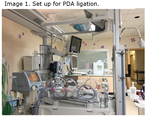
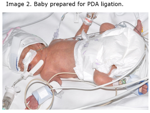
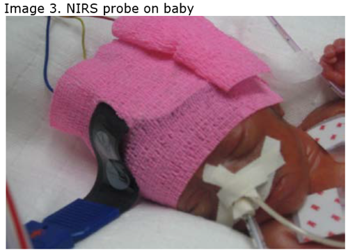
From Scottish data independent risk factors for death by 1 year were FiO2 >40% prior to surgery and lack of prior treatment with cyclo-oxygenase inhibitors (COI). Early post-operative deaths are associated with higher preoperative FiO2 > 60%. Significant complications such as perioperative pneumothorax (2.4%) and vocal cord palsy (4.8%) (26) are infrequent. Internationally reports of Post LIgation Cardiac Syndrome (PLICS) vary from to 30% to 8%. The syndrome is off sever lleft ventricular faiure and poor cardiac output with hypotensin and failing circulation. However the incidence of classic PLIC appears to be falling whilst but post Post LIgation Cardiac-Respiratory Syndrome is probably more common than previously appreciated. This can present with relative hypertension and deteriorating respiratory function 12-48 post operatively with a relative white out on chest X ray. This is also found following catheter closure of the PDA in very preterm infants. The mechanism is almost certainly due to increased systemic vascular resistance and poor diastolic function resulting in pulmonary oedema. For management see Figure 1.

Separate consideration should be given to the more mature infant, > 28 weeks, who are rarely reported in PDA ligation series. Most infants do not require management of the DA. However occasionally an atypical large and persistent DA is present resulting in heart failure often requiring extended periods of invasive or non-invasive respiratory support, failure to thrive and delay in developmental attainment such as sucking feeds. Experience suggests these infants derive considerable clinical benefit from PDA ligation.

PDA ligation referral guidance: a structured approach with robust clinical and echocardiographic assessment

For the reasons described above there are no UK or international guidelines on PDA ligation.

Consideration for ligation depends on robust clinical and echocardiographic assessment (Appendix 1 & 2) based on evolving international standards and timely referral.  

Indications for referral                                                                                                                 

Potential clinical indications for consideration of referral for PDA ligation are listed below. They should be considered in conjunction with a full echocardiographic PDA assessment (Appendix 1) and considered against a clinical and echocardiographic triage score (Appendix 2). Only infants with clinical criteria and a moderate or large PDA, who are unsuited to or who haven’t responded to PGHS2 inhibitors, should proceed to ligation.

  • Persistent systemic hypotension resistant to medical management
  • Significant pulmonary haemorrhage
  • NEC
  • Renal failure or impairment
  • Failure to extubate
  • Cardiac failure not controlled medically (Hiflow/CPAP dependency, failure to thrive)

Echocardiographic assessment of the PDA (Appendix 1)

Echocardiographic assessment of the PDA can be challenging because of the 3D structure of the PDA and variability of shunt depending on clinical support and clinical condition. In addition reporting may also be subject to individual bias and understanding of the correlation of PDA diameter to the patient’s gestation and weight. Therefore it is important to have a robust system of assessment with standardised reports which grade the significance of the PDA.

It is not possible to measure the shunt directly but surrogates can be used looking at PDA diameter and flow patterns, measures of pulmonary blood flow (including LVO or LVO/RVO ratios) and measures of systemic flow. Using these measures PDA can be graded into small, moderate or large shunts (35). Moderate and large shunts can be considered for ligation if the above clinical criteria are met. It is important to recognise that the presence of an atrial shunt in the presence of a PDA can double the anticipated pulmonary blood flow. Atria communication >1mm in association with PDA.1.5mm are associated with increased measures of haemodynamic significance and worse clinical outcomes (37).

Clinical triage scoring system (Appendix 2)

A triage system to determine time to surgical ligation was devised in Toronto (36). Patients must have a moderate or large PDA but triage is based on the illness severity. In Toronto category 1 patients were ligated within 24 hours, category 2 within 3 days and category 3 within 7 days. In Glasgow most cases have been historically category 3 but as we move to look after smaller infants, who may not tolerate medical closure we will need to reconsider this strategy.

Referral process                                                                                                                

Patients should be referred to both the on call Neonatal Consultant at RHC and the on call Consultant Cardiologist. Where the need for PDA ligation is clear, arrangements will be made to admit to NICU as soon as possible (consider against the triage system). Where the need for ligation is uncertain and the referring unit has capability the baby could be presented at JCC remotely. TEAMS consult could also be considered or it may occasionally be possible for a cardiology review to be undertaken at the referring hospital.

Key Points

  • Prolonged PDA exposure is associated with adverse outcomes such as reduced brain MRI volume, heart failure and BPD.
  • Preterm PDA ligation is reserved for infants who have not responded to or who are unsuited to medical treatment (COI or PGHS2 inhibitors)
  • Higher rates of adverse neurodevelopmental outcomes have been linked to PDA ligation but do not persist after risk adjustment.
  • Extubation success post PDA ligation continues to be demonstrated in UK series
  • International centres reporting good outcomes tend to ligate earlier then UK case series but ideally > 21-30 days of life.
  • Post PDA ligation mortality is associated with increasing FiO2 at ligation: < 40% is favourable.
  • Standardised approaches to echocardiographic and illness assessment are essential 
  • Referrals should be (triaged) based on a clinical triage (score) severity score (Appendix 3) where possible.
  • Input by neonatal clinicians is important at JCC
  • Ligation should be undertaken in centres with the ability to support post-operative cardiac and respiratory instability.
Practical process for PDA ligation NICU RHC

On Admission to NICU       

Assessment for ligation

  1. Neonatology assessment
    All infants will be reviewed by a consultant neonatologist and assessed against the clinical and PDA severity scores.

  2. Cardiology assessment
    Formal referral must be made to the on call cardiology team. All infants will be reviewed by a cardiologist have a PDA shunt assessment if not already done (Appendix 2 and 3). Prior to surgery a full paediatric cardiologist structural echo is required. All echocardiographic images and clinical history must be reviewed by a consultant cardiologist and consultant neonatologist with expertise before a final decision to proceed to referral for PDA ligation. The discussions usually take place at the joint cardiac conference (JCC) every Friday but may be earlier if triage indicates urgent surgery may be required.

Patient preparation

  1. Bloods. U/Es, FBC, CRP, X-match of 1 unit of blood. Ensure Hb > 120g/dl. Blood will be requested by the theatre team at the start of the PDA ligation. Blood will be delivered in a sealed cool box and will be returned to blood bank following the procedure if unused. Return of the blood is also the responsibility of the theatre team.
  2. Chest X ray. A recent chest X ray must be available.
  3. Clinical assessment. Ensure that the patient is not septic preoperatively.
  4. Antimicrobial cover. All infants should have perioperative antibiotics. This should be gentamicin, vancomycin and fluconazole unless cultures suggest broader cover is required.
  5. Invasive arterial monitoring. Arterial access, ideally post ductal, is desirable but not essential.
  6. IV access. Two peripheral IV cannulae or a PICC and PVL are required prior to the procedure.
  7. IV fluids. Most infants are on supplemental sodium chloride (oral supplements or breast milk fortifiers). Care should be taken to ensure that IV fluids contain adequate amounts of sodium supplementation. In most cases, a bespoke bag of TPN will be prepared for the baby.
  8. Fasting. Enteral feeds must be stopped 6 hours prior to the procedure (4 hours may be possible if all EBM). Oral supplements and medications may be given up until 4 hours prior to surgery.
  9. Sedation. Commence morphine at 20- 30 microg/kg and consider commencing vecuronium infusion
  10. NIRS and saturations. Application of cerebral NIRS and pre- and post-ductal saturation probes.
  11. Prepare Milrinone for post operative infusion starting at 0.3 microg/kg/min

Preparation for PDA ligation in the NICU (see Appendix 1 for standardised lay out)

Since 2010 all PDA ligations on preterm babies have been undertaken in the NICU. This minimizes handling, the risk of hypothermia and disturbances in ventilation. PDA ligations on term and post-term babies are carried out in theatre.

To ensure a safe surgical environment and minimal disturbance to the infants care it is vital to have the correct equipment and preparation. 

The baby should be ready at least 30 minutes prior to surgery to allow a period of stability.

1. Incubator selection. A decision must be made for every baby referred as to the most appropriate platform for surgery. Where possible this should be the infant’s own incubator and they should be nursed in this incubator on admission to the unit. There are 2 options

  • The Giraffe Omnibed – ideal for all infants regardless of size and humidity requirements. Provides humidity but has the advantage of a removable lid, sides and overhead heater. However resource limited and priority should always be given to the smallest and least mature infant.
  • The Draegar Babytherm. This is suitable for infants > 1.8kg and who no longer require humidity.

2. Incubator orientation. The incubator must be placed in a reverse position with the foot end at the wall (medical gas supply end). The baby should be positioned with their head at the incubator foot (at the gas supply end). This facilitates access for the anaesthetic team to the baby’s head and the gas supply. Appendix 2

3. Incubator Position in the unit. PDA ligation should always be undertaken in the same cot position in any one of the four bed bays. In practice this is always on the right hand side, nearest the window.

4. Equipment for surgical team from theatres

  • Blue trolley with disposal bag available
  • Plug bank on blue trolley

Imaging and surgical approach.

Prior to ligation, it is important that cardiology ascertain the correct anatomy, whether the baby has a left or right arch and the position of the duct. This is typically done by echo although in unusual cases a CT may be required. The usual approach is left thoracotomy. Occasionally median sternotomy can be carried out if baby is intolerant of left position. Very occasionally a right thoracotomy may be required

NICU team prior to theatre team arrival

  1. Ensure surgical room is free of parents and is quiet
  2. Prepare surrounding space as per Appendix 2 and Image 1 and 2.
    • Pull incubator foot away from wall to allow access for anaesthetist at the foot of the incubator (baby’s head)
    • Infusion pumps will be at wall behind the anaesthetist.
    • Ventilator and monitors should be placed to the right of the anaesthetist
    • Ensure air oxygen blender and rebreathing circuit available.
    • Ensure clear space available to the left (surgeon, diathermy, surgical light, sterile trolley and theatre staff)
    • Ensure clear space available to the right (surgical assistant, anaesthetist’s trolley and rubbish bag)
  1. Prepare the baby as per Image 2 and Image 3 ensuring pre and post ductal probes,  cerebral NIRS, ECG leads of the hemithorax
    • *Place on warm “transwarmer” wrapped in single sheet if procedure in Babytherm but not Giraffe Omnibed. Cover baby in bubble wrap, remove incubator lid and sides and turn on overhead heater on arrival of the theatre team.
    • Complete a theatre anaesthetic check list as if the patient is going to theatre
    • Transfer care to theatre team but baby’s nurse remains in the clinical area.

Surgical Team

Surgical pause and provision of the following is the responsibility of the surgical team

  • Diathermy unit
  • Theatre packs
  • Surgical light
  • Theatre resuscitation / intubation trolley
  • Anaesthetic drugs.

Operative care                                                                                                                    

Although the care of the baby is the responsibility of the theatre team during surgery the following are encouraged:

  • Neonatal nurse remains present throughout to advise on any difficulties with monitoring, pumps or practical management.
  • TPN continues throughout procedure
  • Vancomycin infusion continues throughout the procedure

Post-operative care

See Image 1 for guidance.                                                                                               

Potential post-operative complications include pneumothorax (very rare), acute early hypotension( 1st 6 hours), later hypotension and circulatory insufficiency(PLIC) and transiently worsening cardiac failure and respiratory instability (PLICR) due to rapid rises in systemic vascular resistance (8) best treated by inodilators (32,33,34). We recommend commencing Milrinone in all infants at 0.3/kg/min immediately post surgery.  

Attention should be paid to BP and NIRS measurements. Cardiac echo should be undertaken if there is hypotension or hypoxia is severe to review function, to ensure clip position and exclude pericardial effusion.  

Routine post-operative care includes the following

  • Chest X ray immediately post procedure
  • Blood gas immediately post procedure
  • Continue antibiotics for 24 hours
  • Cardiac echo in the first (1st) 24 hours following the procedure (earlier if clinical instability)
  • Ensure adequate analgesia – morphine infusion of 20-30 micrograms/kg/hour
  • Recommence feeds at consultant neonatologist’s discretion – usually within a few hours
  • Chest drain removed at consultant neonatologist’s or cardiac surgeon’s discretion – usually the following day.
  • Formal cardiac echo prior to discharge.

Potential complications

  • Acute blood loss and hypovolemia
  • Post Ligation Impaired LV contractility +/- hypotension (PLiCS)
  • Post ligation cardio-respiratory syndrome (PLiCRS
  • Pneumothorax – not small residual post-operative air collection.
  • Injury to left (RLN) recurrent laryngeal nerve causing vocal cord palsy – but not detectable in intubated patients
  • Incomplete duct ligation – persistent flow
  • Ligation of wrong vessel (left pulmonary artery/ aorta)
  • Unmasking of coarctation of the aorta
  • Compression of the left main bronchus.
  • Chylothorax

Although there is risk of left sided vocal cord paralysis after PDA ligation we do not routinely perform MLB. However this should be considered in babies with persisting stridor, worsening BPD and/ or feeding problems after extubation following PDA ligation (17). Most recover spontaneously.

Management of post-operative hypotension or Post LIgation Cardiac Syndrome (PLIC)  

  1. Ensure all infants on low dose milrinone at (0.3 micrograms/kg/min)
  2. Ensure adequately sedated and consider muscle relaxation if difficulties with oxygenation.
  3. Immediate post op hypotension give Volume: 20ml/kg 0.9% NaCl given over 20 minutes or transfuse as packed red cells if Hb < 14g/dl. Consider Hydrocortisone (2.5mg/kg 6-12 hourly).
  4. Late hypotension add adrenaline
  5. Late respiratory deterioration and adequate BP increase milrinone up to 0.7 micrograms/kg/min and consider diuretics.

Follow up after PDA ligation

Infants referred for PDA ligation are at high risk of BPD and neurodevelopmental disability. All infants should have formal neurodevelopmental assessment until at least 2 years of age.

Images

Appendix 1: Schematic diagram for PDA ligation

Appendix 2: Standardised PDA assessment

Appendix 3: Clinical Triage Score

From: EL-Khuffash, Afif. (2014). Neonatal Echocardiography Teaching Manual.

References
  1. Brown ER. Increased risk of bronchopulmonary dysplasia in infants with patent ductus arteriosus. J Pediatr. 1979;95:865-6.
  2. Kitterman JA, Edmunds LH Jn, Gregory GA, Heymann MA, et al. Patent ductus arteriosus in premature infants: incidence and relation to pulmonary disease and management. N Eng J Med 1972;287:473
  3. Clyman R, Narayanan M. Patent ductus arteriosus: a physiologic basis for current treatment practices. Current Topics in Neonatology. Philadelphia: WB Saunders; 2000:71-91
  4. Lemmers PM, Toet MC, Van Bel F. Impact of patent ductus arteriosus and subsequent therapy with indomethacin on cerebral oxygenation in preterm infants. Pediatrics 2008;121:142-147.
  1. Gerhardt T, Bancalari E: Lung Compliance in Newborns with Patent Ductus Arteriosus before and after Surgical Ligation. Neonatology 1980;38:96-105.
  2. Balsan, M.J, Jones, J.G. and Guthrie, R.D. (1991), Effects of a clinically detectable PDA on pulmonary mechanics measures in VLBW infants with RDS. Pediatr. Pulmonol., 11: 161-165.
  3. Cotton RB, Stahlman MT, Bender HW, Graham TP, Catterton WZ, Kovar I. Randomized trial of early closure of symptomatic patent ductus arteriosus in small preterm infants. J Pediatr. 1978 Oct;93(4):647-51
  4. Laughon M, Bose C, Benitz WE. Patent Ductus Arteriosus Management: What Are the Next Steps? J Pediatr 2010;157(3):355-7.
  5. Mitra S, Scrivens A, von Kursell AM, Disher T. Early treatment versus expectant management of hemodynamically significant patent ductus arteriosus for preterm infants. Cochrane Database of Systematic Reviews 2020, Issue 12. Art. No.: CD013278.
  6. Evans_P, O'Reilly_D, Flyer_JN, Soll_R, Mitra_S. Indomethacin for symptomatic patent ductus arteriosus in preterm infants.Cochrane Database of Systematic Reviews 2021, Issue 1. Art. No.: CD013133.
  7. Liebowitz M, Koo J, Wickremasinghe A, Allen IE, Clyman RI. Effects of Prophylactic Indomethacin on Vasopressor-Dependent Hypotension in Extremely Preterm Infants. J Pediatr. 2017 Mar;182:21-27
  8. Liebowitz M, Clyman RI. Prophylactic Indomethacin Compared with Delayed Conservative Management of the Patent Ductus Arteriosus in Extremely Preterm Infants: Effects on Neonatal Outcomes. J Pediatr. 2017 Aug;187:119-126.
  9. Ment LR, Vohr B, Allan W, Westerveld M, Sparrow SS, Schneider KC, Katz KH, Duncan CC, Makuch RW. Outcome of children in the indomethacin intraventricular hemorrhage prevention trial. Pediatrics. 2000 Mar;105(3 Pt 1):485-91
  10. Lemmers PM, Benders MJ, D'Ascenzo R, Zethof J, Alderliesten T, Kersbergen KJ, Isgum I, de Vries LS, Groenendaal F, van Bel F. Patent Ductus Arteriosus and Brain Volume. Pediatrics. 2016 Apr;137(4):e20153090. doi: 10.1542/peds.2015-3090. Epub 2016 Mar 30. PMID: 27030421
  11. El-Khuffash A, Rios DR, McNamara PJ. Toward a Rational Approach to Patent Ductus Arteriosus Trials: Selecting the Population of Interest. J Pediatr. 2021 Jun;233:11-13. doi: 10.1016/j.jpeds.2021.01.012.
  12. Liebowitz M, Katheria A, Sauberan J, et al. Lack of Equipoise in the PDA-TOLERATE Trial: A Comparison of Eligible Infants Enrolled in the Trial and Those Treated Outside the Trial. The Journal of Pediatrics. 2019 Oct;213:222-226.e2.
  13. Clyman RI, Kaempf J, Liebowitz M, Erdeve O, Bulbul A, Håkansson S, Lindqvist J, Farooqi A, Katheria A, Sauberan J, Singh J, Nelson K, Wickremasinghe A, Dong L, Hassinger DC, Aucott SW, Hayashi M, Heuchan AM, Carey WA, Derrick M, Fernandez E, Sankar M, Leone T, Perez J, Serize A; PDA-TOLERATE Trial Investigators. Prolonged Tracheal Intubation and the Association Between Patent Ductus Arteriosus and Bronchopulmonary Dysplasia: A Secondary Analysis of the PDA-TOLERATE trial. J Pediatr. 2021 Feb;229:283-288
  14. Clyman, R.I., Hills, N.K., Cambonie, G. et al.Patent ductus arteriosus, tracheal ventilation, and the risk of bronchopulmonary dysplasia. Pediatr Res(2021). 
  15. Bussmann N, Smith A, Breatnach CR, McCallion N, Cleary B, Franklin O, McNamara PJ, El-Khuffash A. Patent ductus arteriosus shunt elimination results in a reduction in adverse outcomes: a post hoc analysis of the PDA RCT cohort. J Perinatol. 2021 May;41(5):1134-1141.
  16. Gersony WM, Peckham GJ, Ellison RC, Miettinen OS, Nadas AS. Effects of Indomethacin in premature infants with patent ductus arteriosus: results of a national collaborative study. J Pediatr 1983;102:895-906 
  17. Waleh N, McCurnin DC, Yoder BA, Shaul PW, Clyman RI. Patent ductus arteriosus ligation alters pulmonary gene expression in preterm baboons. Pediatr Res. 2011 Mar;69(3):212-6.
  18. Cassady G, Crouse DT, Kirklin JW et al. A randomized, controlled trial of very early prophylactic ligation of the ductus arteriosus in babies who weighed 1000 g or less at birth. N Engl J Med 1989;320:1511-6.
  19. Clyman R, Cassady G, Kirklin JK, Collins M, Philips JB 3rd. The role of patent ductus arteriosus ligation in bronchopulmonary dysplasia: reexamining a randomized controlled trial. J Pediatr. 2009 Jun;154(6):873-6 … prophylactic ligation is bad for BPD but not NEC
  20. Kabra NS, Schmidt B, Roberts RS, et al. Neurosensory impairment after surgical closure of patent ductus arteriosus in extremely low birth weight infants: results from the Trial of Indomethacin Prophylaxis in Preterms. J Pediatr 2007;150:229–34
  21. Weisz DE, Mirea L, Resende MHF, Ly L, Church PT, Kelly E, Kim SJ, Jain A, McNamara PJ, Shah PS. Outcomes of Surgical Ligation after Unsuccessful Pharmacotherapy for Patent Ductus Arteriosus in Neonates Born Extremely Preterm. J Pediatr. 2018 Apr;195:292-296.
  22. Heuchan AM, Hunter L, Young D. Outcomes following the surgical ligation of the patent ductus arteriosus in premature infants in Scotland. Arch Dis Child Fetal Neonatal 2012;97:F39-44. doi:10.1136/adc.2010.206052
  23. Warnock, A., Szatkowski, L., Lakshmanan, A. et al.Surgical management of patent ductus arteriosus in pre-term infants - a british paediatric surveillance study. BMC Pediatr21, 270 (2021).
  24. Lemmers PM, Molenschot MC, Evens J, Toet MC, van Bel F. Is cerebral oxygen supply compromised in preterm infants undergoing surgical closure for patent ductus arteriosus? Arch Dis Child Fetal Neonatal Ed. 2010 Nov;95(6):F429-34.
  25. Brooks JM, Travadi JN, Patole SK et al. Is surgical ligation of patent ductus arteriosus necessary? The Western Australian experience of conservative management. Arch Dis Child Fetal Neonatal Ed 2005;90:F235-9.
  26. Lee, J.H., Lee, H.J., Park, HK. et al.Surgical ligation of patent ductus arteriosus in preterm neonates weighing less than 1500g: a 9-year single center experience. J Cardiothorac Surg15, 144 (2020). https://doi.org/10.1186/s13019-020-01191-2
  27. Jhaveri N, Moon-Grady A, Clyman RI. Early surgical ligation versus a conservative approach for management of the patent ductus arteriosus that fails to constrict after indomethacin treatment. J Pediatr 2010;157:381-7.
  28. Teixeira LS, Shivananda SP, Stephens D et al. Postoperative cardiorespiratory instability following ligation of the preterm ductus arteriosus is related for early need for intervention. J Perinatology 2008;28:803-10.
  29. El-Khuffash AF, Jain A, Weisz D, Mertens L, McNamara PJ. Assessment and treatment of post patent ductus arteriosus ligation syndrome. J Pediatr. 2014 Jul;165(1)
  30. Clyman RI, Wickremasinghe A, Merritt TA, et al. Hypotension following patent ductus arteriosus ligation: the role of adrenal hormones. The Journal of Pediatrics. 2014 Jun;164(6)
  31. McNamara PJ, Sehgal A. Towards rational management of the patent ductus arteriosus: the need for disease staging. Arch Dis Child Fetal Neonatal Ed. 2007;92:F424-F427
  32. EL-Khuffash, Afif. (2014). Neonatal Echocardiography Teaching Manual.
  33. Rios D, Martins F, EL-Khuffash A, Weisz D, Giesinger R, Mcnamara P. (2020). Early Role of the Atrial-Level Communication in Premature Infants with Patent Ductus Arteriosus. Journal of the American Society of Echocardiography. 2020; 34(4).
  34. Weisz DE, Mirea L, Rosenberg E, Jang M, Ly L, Church PT, Kelly E, Kim SJ, Jain A, McNamara PJ, Shah PS. Association of Patent Ductus Arteriosus Ligation With Death or Neurodevelopmental Impairment Among Extremely Preterm Infants. JAMA Pediatr. 2017 May 1;171(5):443-449. doi: 10.1001/jamapediatrics.2016.5143. PMID: 28264088; PMCID: PMC5470355.
Editorial Information

Last reviewed: 22 October 2022

Next review: 01 October 2025

Author(s): Dr Anne Marie Heuchan – Neonatal Consultant RHC & Dr A MacLaren – Neonatal consultant RHC

Co-Author(s): Other Professionals Consulted: Cardiologists (RHC), Cardiac Surgeons (RHC), Cardiac Anaesthetist (RHC), Neonatologists (RHC), SNHPG executive group