Seldinger chest drain insertion and management
exp date isn't null, but text field is
Objectives
This guidance is for Medical staff and Advanced Neonatal Nurse Practitioners working in Neonatal Intensive Care settings within the MCN for Neonatology, West of Scotland. It relates to the insertion of the Fuhrman Pleural/Pericardial chest drain.
The existence of this guideline does not preclude the use of other chest drain methods which may be preferred for certain circumstances.
The Seldinger method of chest drain insertion has rapidly become the preferred technique for most neonatologists. This is because it is viewed as less “invasive” than the previously used trocar method, and with practice can be inserted quickly and safely. As with any procedure however it carries risks and as such appropriate preparation (including case selection) and technique are vital.
- Pneumothorax
- Pleural effusion/chylothorax
For the Seldinger technique to be safe it is vital that there is a sufficiently large collection of air or fluid in the pleural space for insertion of the needle and so separate needle thoracocentesis prior to drainage should be avoided except in extremis. Small accumulations of pleural air or fluid which the baby is tolerating clinically should be managed conservatively,
Where there is leakage of fluid from a previous drain site (i.e. from pleural effusions /chylothoraces) the Seldinger method should not be used unless an adequate residual fluid collection has been demonstrated. An alternative drain that does not rely on initial needle thoracocentesis should be used.
A chest drain is indicated when there is significant respiratory or circulatory compromise. The decision to proceed to chest drain insertion will generally be made after discussion with the appropriate consultant and following review of current X-rays or other imaging such as thoracic USS. The procedure should be performed by an experienced clinician or under the direct supervision of a consultant, supported, where possible, by a skilled assistant.
- Frequently time pressure will not allow full discussion with parents prior to insertion of a chest drain, in which case a full discussion should be had at the first opportunity subsequently. This discussion should be clearly documented in the patient’s notes.
- Review analgesia/sedation for the baby – consider a morphine or fentanyl bolus. Oral sucrose/dextrose can be used. Lidocaine should be administered subcutaneously prior to drain insertion.
- Organise equipment and brief assistant.
- Sterile dressing pack, gloves, gown and appropriate skin sterilising preparation
- Lidocaine 1% with syringe and needles for drawing up and injection (allow for up to 0.3ml/kg)
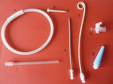
- Fuhrman pleural drainage set, Fig. 1 (Cook product G03974) which includes J tip guide wire in its plastic sheath (a) with introducer (b), access needle (c), dilator (d), pigtail catheter (e), 3 way tap (f) and tubing adapter (g).
NB for connection to underwater drainage systems with a male connector, a separate adaptor will be required - A needle free adaptor device (e.g. Vadsite/bionector/smartsite etc) to put on the side port of the three way tap.
- “Tegaderm” (or similar occlusive dressing) for fixing catheter to the skin. Sutures to secure the drain may be considered if there are concerns about the catheter being fixed with the fixation device/Tegaderm, e.g. where there is fluid leak from around the drain or concerns regarding skin integrity around the insertion site.
- Drainage tubing and underwater seal chest drain bottle (See underwater drainage instructions below)
- Low regulator suction system
Fig 1.
- Position baby with affected side raised and identify site – anterior axillary line, 4th to 6th intercostal space. The nipple usually lies in the 5th intercostal space and can be used as a landmark in neonates. The drain must not be inserted through the nipple
- Prepare for aseptic procedure including sterilisation of skin as per current policy.
- Infiltrate skin and proposed track with lidocaine 1% - up to 0.3ml/kg
- Guide the access needle through the chest wall while aspirating with attached syringe. It is very important to guard against over insertion by gripping the access needle between thumb and index finger 1cm from its tip, Fig. 3. Stop and hold position as soon as air or fluid can be aspirated. Do not aspirate any more that is necessary to ascertain that the needle is in the pleural cavity as this may increase the risk of lung injury.
- Insert guide wire into needle with the help of the white plastic introducer. Watch for the mark on the wire which indicates adequate level of insertion when it is just entering the hub of the needle, Fig 4. If resistance is felt when inserting guide wire stop and remove wire and needle.
- While maintaining the wire in position, remove the needle.
- While maintaining the wire in position, introduce dilator over the wire and dilate the entry track with a gentle twisting motion. NB – it is only necessary to insert the dilator until the widest point of the tapered end passes through the skin. This can be assured by holding the dilator about 1.5 cm from the tip, thus avoiding any deeper insertion. Remove the dilator.
- While maintaining the wire in position, feed the pigtail catheter over the wire and through the entry track with a gentle twisting motion. When the catheter has been inserted 1-2cm beyond the drainage holes the guide wire can be removed
- Fix in place with occlusive dressings (“Tegaderm”) taking care to avoid kinking at the point of entry to the chest wall. Occlusive dressings alone may be insufficient to secure chest drains where there is leaking pleural, in this situation suturing in place should be considered.
- Luer lock the 3 way tap to the catheter and attach, in-line, to tubing adapter and drainage system. Attach a needle free adaptor to the third arm of the tap for active aspiration of air or fluid if necessary.
In the event that an infant requires a further chest drain because the initial one has been removed or has become dislodged it is not recommended to insert a new drain via the same entry site. This is because an internal tract may have formed which will increase the chance of the wire taking an inappropriate route within the thoracic cavity, eg towards the heart. When multiple drains are required, however, small size may pose difficulties in finding a new entry point for each drain and so clinical judgement must be used in deciding where to place a new drain.
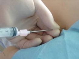 Fig 3. Fig 3. |
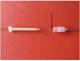 Fig 4. Fig 4. |
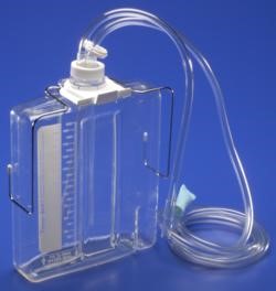 Fig 5. |
|
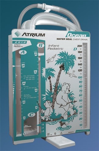 Fig 6. |
|
- X-Ray after insertion of drain to establish position
- Repeat X-Ray as clinically indicated
- Do not X-Ray routinely after removal of drain. This should be guided by clinical factors.
- Document procedure in medical notes and in appropriate data management system
- Record condition of drain site and functioning of drain with routine hourly observations of the baby
- If fluid is drained, monitor and record fluid volume collected hourly in the drainage chamber
- When the chest drain has stopped draining, the water seal will no longer be bubbling. The drain should be clamped for a few hours before removal to ensure there is no reacummulation of fluid or air. If, after 2-4 hours, the infant remains stable, the drain can be removed.
- While taking sterile precautions, pull the catheter from its site, taking care of splatter from the pigtail as it springs out.
- Apply steristrip and a sterile dressing; suturing of the wound is not required.
- The tip of the pigtail catheter can be sent for culture if indicated
- Haemorrhage of subcostal vascular bundle – reduce risk by placing the drain just above a rib rather than below
- Damage to thoracic structures during needle/guide wire insertion – avoid inserting a drain for small air/fluid accumulations and do not carry out needle thoracocentesis before inserting drain if this can be avoided.
- Scarring – do not use purse string suture to fix drain in place
- Infection – use sterile technique with a helper to keep control of the flexible guide wire within the sterile field. The use of antibiotic prophylaxis is not routine.
- In the event that the operator considers that the heart or liver (or other organ) may have been perforated by the drain the 3 way tap should be immediately turned ‘off to the baby’ and the drain left in situ. IT IS VITAL THAT THE DRAIN IS NOT REMOVED AT THIS POINT.
- The consultant, if not already present, should be contacted and asked to attend. Emergency chest X-ray should be performed; ultrasound imaging to identify the drain site may be helpful if a skilled operator is available. Appropriate stabilisation of vital parameters should be attempted, though in the case of a cardiac injury this may only be possible with surgery. If necessary, there should be immediate consultation with the cardio-thoracic team (cardiac injury) or general surgical team (liver injury) and emergency transfer organised.
Last reviewed: 03 October 2022
Next review: 01 October 2025
Author(s): Allan Jackson – Consultant neonatologist PRM
Approved By: WoS Neonatology MCN

